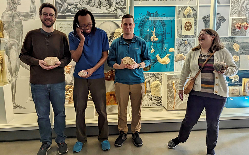
Late last month a cadre of students from the Schar School of Policy and Government’s biodefense graduate program visited the National Museum of Health in Medicine in Silver Spring, Maryland, not far from the George Mason University campuses.
The 150-year-old museum is known for its collections that depict the human anatomy and everything that can befall it, in sometimes stark and gory detail. What else would you expect from a museum with a pathology guide with listings for “bilateral nephrolithiasis (kidney stones),” “liver, hydatid cyst from tape worm,” and an all-time-favorite, “trichobezoar (human hairball from stomach).”
Taking biodefense students who study in a program that bridges the gap between science and policy to a medical museum was meant to “explore former medicinal interventions, medical procedures such as facial reconstructive surgery and amputations, and infections such as gangrene and pneumonia dating back to the Civil War era,” said Lauren E. Quattrochi, the pharmacologist, virologist, electrophysiologist, and adjunct professor in the biodefense program best known as “Dr. Q.”
“This glimpse back in history shows how far we've come to treatments on the battlefield, and what threats they faced in the past,” she said, “including sulfur mustard gas in World War I versus what they could encounter now. We’ve made great strides in our medical countermeasures resilience but there are always new and evolving threats that we must be ready to defeat.”
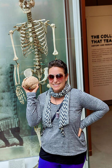
Not only did the students see some of the 3 million plasticized, petrified, and preserved artifacts in the museum’s collection, but they were offered opportunities to handle them. Other than the occasional squeamish response, the trip fulfilled its educational purpose.
“It was surreal to see and hold a heart,” said PhD student and graduate research assistant Becca Earnhardt. “For me, it really drove home that these cases we read about in class are not just another statistic or casualty.”
Brain and nervous system models were of particular interest, she said, and the museum did not disappoint.
“The museum has an extensive collection of brain and nervous system models, including samples showing signs of hemorrhaging and other traumatic brain injuries,” Earnhardt said. “One of the key discussions we had as a class was about the connectome,” which, she explained, is a comprehensive mapping of the human brain, including the nerves and synapses.
“An element that is of great interest in biodefense is how the connectome can inform medical countermeasures development and treatments for nerve agent exposure,” she said.
The students were guided through exhibits depicting the impact of infectious diseases on the human body, including liver and kidney specimens displaying the effects of a viral hemorrhagic fever infection.
“Seeing the impact of these deadly diseases up close truly highlights how important it is to find novel treatments,” Earnhardt said. “We were also able to view samples displaying the effects of smallpox infection, an eradicated disease. Most people don’t know what the signs and symptoms of the disease look like. Because smallpox poses a major threat to human health if it reemerged, it was important for us to see the consequences of an infection.”